
What Is a Cone Beam CT Scanner?
A cone beam CT scanner is a specialized digital x-ray unit that allows 3 dimensional imaging of the structures of the mouth, jaws and face. Cone beam images allow us to accurately visualize your specific anatomy at extremely high resolution and in several different viewing planes. 3D films yield a wealth of information that is used to precisely plan your treatment before any procedures begin.
A cone beam CT scan can show the exact position of impacted and malpositioned teeth and will enable us to treatment plan your procedure based on your exact anatomy.
Why Do We Have a Cone Beam CT Scanner?
We have invested in this technology to better serve our patients and to provide the peace of mind that our diagnosis and treatment plans are based on the most comprehensive information found in dentistry and medicine today. It has always been the goal of Lakewood Oral and Maxillofacial Surgery Specialists to combine modern surgical practices and outstanding patient care with state of the art facilities. Our in-house cone beam CT scanner allows us to provide for our patients the highest level of diagnosis and patient care possible.

Why Use a Cone Beam CT Scanner?
Lakewood Oral and Maxillofacial Surgery Specialists strongly believe that better planning leads to more predictable treatment results. These state-of the-art scans are accomplished in a matter of seconds and allow us to identify and accurately measure key anatomical structures such as the exact location of nerves, blood vessels, sinuses, etc.
A cone beam CT scanner is specific to the mouth, jaws and face.
A cone beam CT scanner enables us to obtain clearer and more specific images of teeth, jaws and associated structures.
A cone beam CT scanner is more efficient.
Taking a 3D image in our office is a simple process. The scan only takes 9 seconds and in less than a minute the images will be available on the computer to view. Once you view your film, you will truly appreciate the 3D images and it will allow you to truly visualize your anatomy and better understand your treatment options.
A cone beam scanner has nowhere near the radiation exposure of a traditional hospital CT scanner.
Understanding that many patients are concerned about radiation exposure, we have chosen a system with a very low radiation output. A cone beam CT scan uses at least 10 times less radiation than traditional medical CT scans.
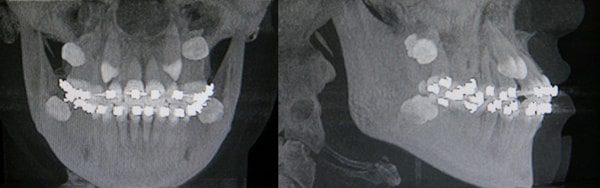
The cone beam CT scanner allows us to see the exact position of impacted teeth.

The above patient has numerous impacted maxillary and mandibular teeth.

The cone beam CT gives us the opportunity to view structures (like impacted teeth) in multiple different planes in order to visualize your exact anatomy.
Cone beam CT scans allow us to identify the exact position of nerves and blood vessels surrounding impacted teeth.

The information collected from the cone beam CT scanner allows to see the exact position of nerves and blood vessels (red).
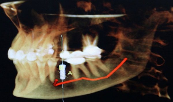
This 3D reconstructive image allows us to precisely plan dental implant procedures. Note that the nerve is highlighted in red
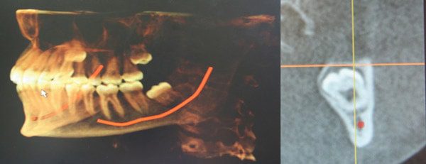
The information collected from the cone beam CT scanner allows to see the exact position of nerves and blood vessels (red).
Exact implant placement can be achieved while avoiding vital structures. Note the position of the nerve in red.
Cone beam CT scans allow us to precisely and accurately place dental implants.
For patients missing single teeth (anterior or posterior), multiple teeth or all of their teeth, the cone beam CT scanner allows us to accurately and predictably work-up implant cases and place implants with extreme precision and accuracy. Note the above implant work-up for a patient missing all lower teeth versus the actual placement of the implants in the postoperative film.

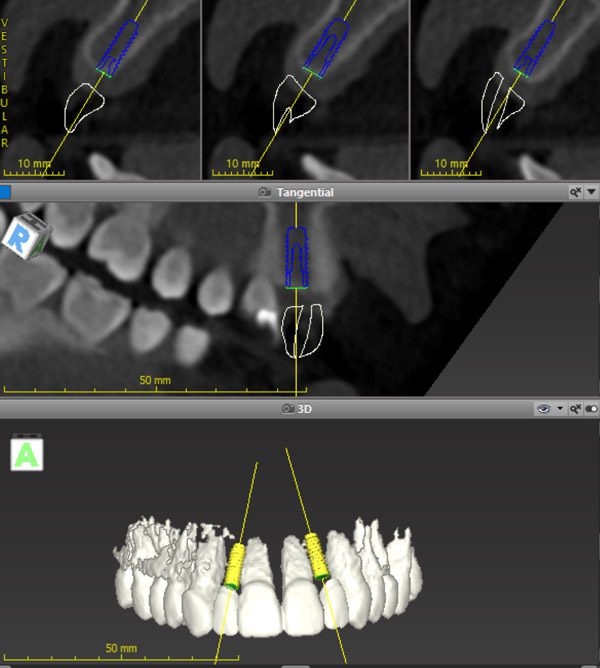
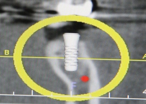
Dental implant treatment planning using the latest computer software and the cone beam CT scanner. Both of the planned implants above are planned with the position of the final crown so that aesthetics are ideal.

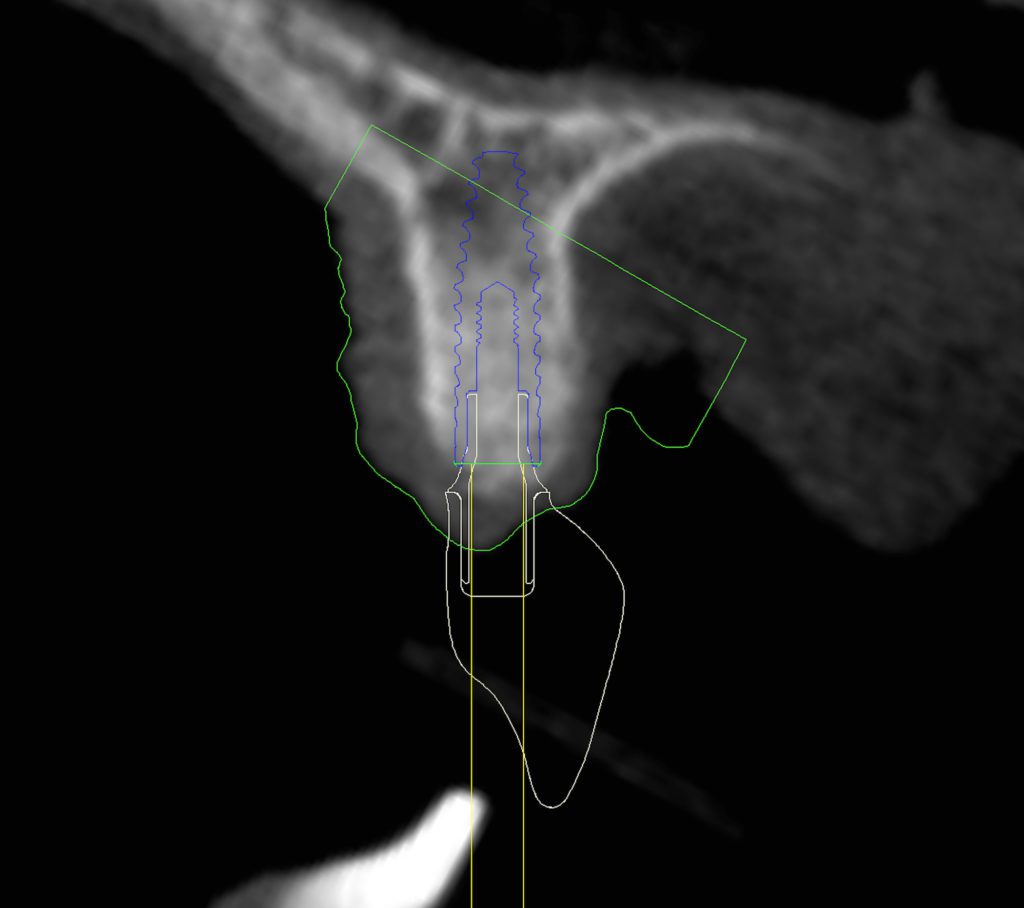
Note the accuracy of the planned dental implant in the figure on the right vs. the final position of the actual dental implant on the left.


